Scintigraphy of the bones
Skeletal scintigraphy (also bone scintigraphy) is a nuclear medical examination method. This involves the use of very low levels of radioactive radiation for a short period of time to monitor bone metabolism. Thus, various diagnoses can be made. How this works exactly, what to consider before and after a bone scintigraphy…
approx. 3-4 hrs.
Duration of examination
approx. 1 liter
Drinking between injection and late shots
Where can you have a bone scintigraphy performed in Munich?
Skeletal scintigraphy is particularly well suited for detecting changes in bone metabolism. This facilitates tumor diagnosis for both bone tumors and other cancers. Especially for patients with lung cancer, breast cancer, kidney cancer or prostate cancer, bone scintigraphy supports the accompanying diagnostics. Our specialists will discuss your needs with you personally and individually.
When is a bone scintigraphy useful?
Bone scintigraphy is used whenever increased activity of bone metabolism is present or suspected.
In case of (suspected) cancer
Skeletal scintigraphy may be helpful for various types of cancer. Tumors often form metastases (metastases or daughter tumors) in the bone. This may be the case, for example, with lung cancer, breast cancer or prostate cancer. Bone scintigraphy also provides valuable information if bone cancer is suspected.
For inflammation
Inflammation of the joints (arthritis) as well as inflammation of the bones and bone marrow lead to a significantly increased bone metabolism. Bone scintigraphy is used, for example, to locate foci of inflammation. Prior to radiosynoviorthesis (joint therapy with radioactive substances), the inflammation in the joint is first detected using skeletal scintigraphy.
For bone injuries
Bone metabolism may be elevated for a variety of reasons. Injuries to certain bone components or malfunctions of the autonomic nervous system bring about increased metabolism. Also a reason could be the healing of a recent bone fracture – in the healing phase the metabolic activity is typically significantly increased.
How does a bone scintigraphy work?
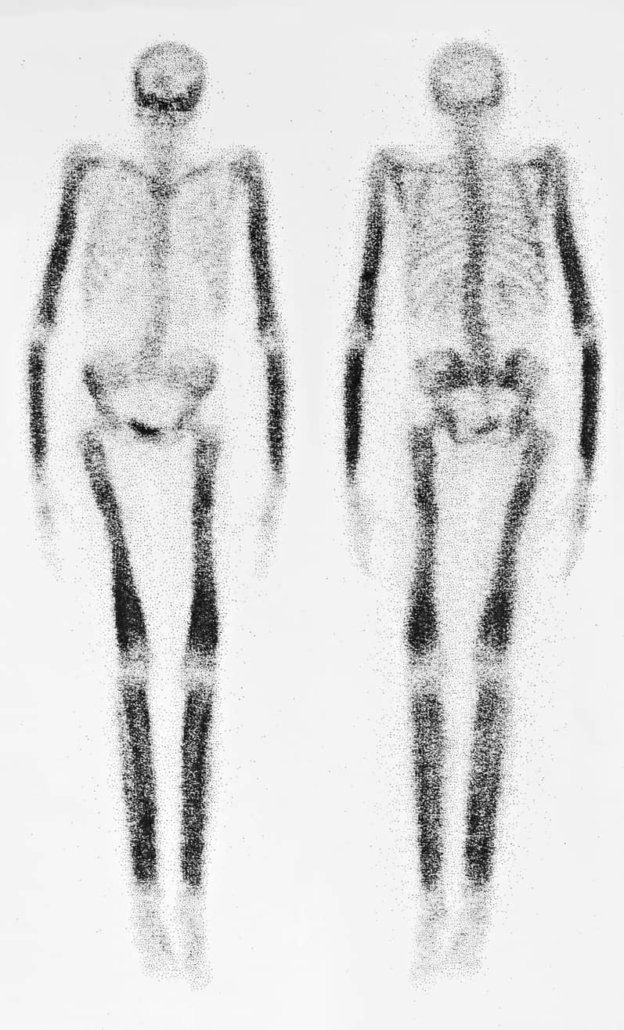
In a bone scintigraphy, a weakly radioactive substance (radiopharmaceutical) is first injected into a vein in the arm. The radiopharmaceutical spreads throughout the body with the blood and then accumulates in the bone. The amount of accumulated agent provides information about the metabolic activity in the different areas of the skeleton is.
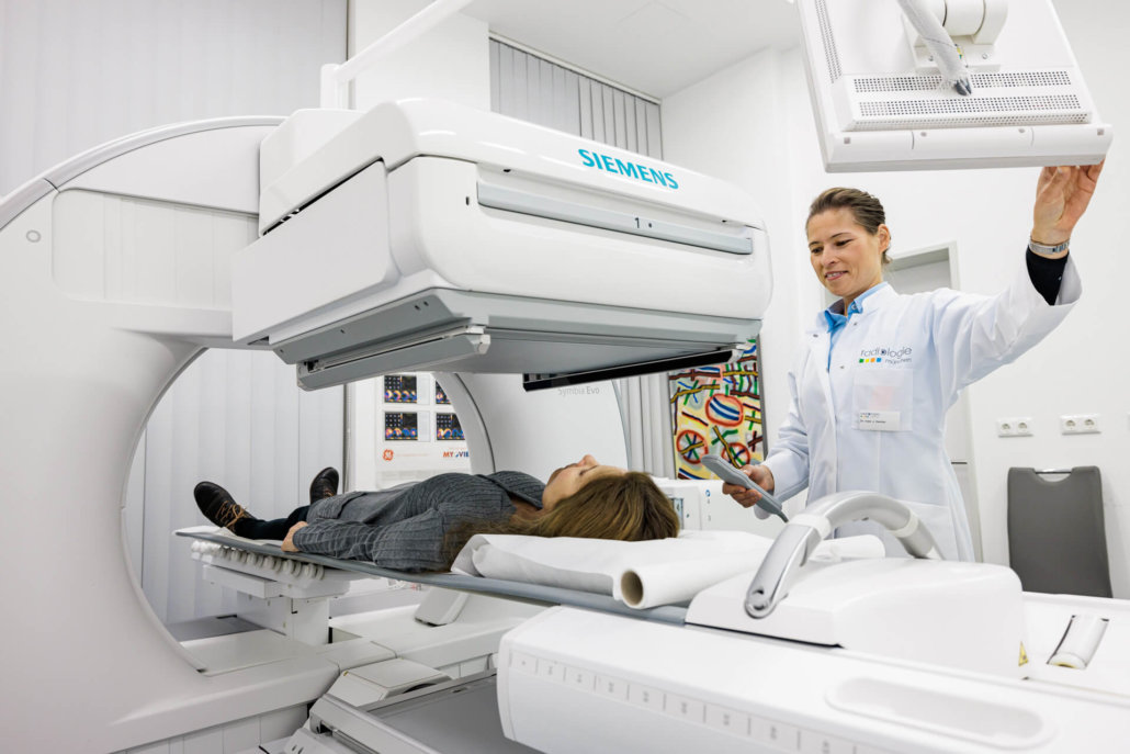
Nuclear physicians can measure the gamma radiation emitted by the radiopharmaceutical with a so-called gamma camera. The data collected in this way can be assembled by a computer into an image of bone metabolism, the scintigram.
What are the advantages of bone scintigraphy?
Like any other nuclear medicine examination method, bone scintigraphy is a non-invasive examination method. This means that the patient’s body is not injured for the examination. Because the radioactive radiation is very low and lasts only a short time, skeletal scintigraphy can be used in a very large number of patients.
In contrast to an X-ray examination, skeletal scintigraphy images pathological changes and processes at a very early stage. For this reason, doctors like to use them to detect disease foci in the bone system at a very early stage.
Which radiopharmaceutical is used?
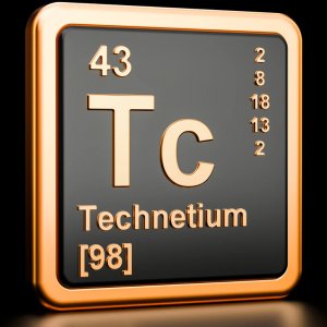
Scintigraphy of bone is performed to mark the increased phosphate content in bone. This is done by a weakly radioactive agent with the ingredient 99mTc. Tc stands for the chemical element technetium. Technetium accumulates more in the phosphate-rich areas (i.e., the diseased parts of the bone) and then becomes visible on the images as a dark area.
What is the procedure for a bone scintigraphy at Radiologie München?
A scintigraphy takes approximately 3 to 4 hours. You do not have to be sober.
The test substance is injected into the arm vein. For certain questions (e.g. question about arthritis), we may take early images for which they lie on the gamma camera for about 10-15 minutes. Here, the so-called blood pool or soft tissue phase is prepared, which provides us with information about increased distribution in the soft tissue as is the case with inflammation. Now it’s time for you to wait and drink – at least one liter before the pictures are taken. This measure serves to ensure that the unbound activity on the bone is increasingly excreted via the kidneys in order to improve image quality and reduce radiation exposure.
As a rule, the images are then taken on the gamma camera after about 3 hours. First, the whole skeleton is recorded. You should lie as still as possible to avoid blurred images. Based on this image, a decision is made as to whether additional images of specific body parts should be taken.
What are the risks and side effects of bone scintigraphy?
Since the amount of radioactive substance used is extremely small, more severe allergic reactions almost never occur during skeletal scintigraphy. Very rarely, mild skin rash or itching may occur. Radiation exposure is also extremely low. It is usually lower than that of a computed tomography scan.
Pregnant women are examined with the help of skeletal scintigraphy only in emergencies, as is the case with other examination methods using radioactive radiation. Breastfeeding mothers should not normally breastfeed their child for 48 hours and should be sure to consult with their physician prior to the examination.
