Myocardial scintigraphy – gentle, safe
Cardiac scintigraphy (myocardial scintigraphy) is a gentle examination method which, as a non-invasive examination procedure (in contrast to cardiac catheterisation), maps the blood flow to your heart muscle using low levels of radioactive radiation. With this examination, circulatory disorders (ischaemia) can be detected at an early stage and pre-existing coronary heart disease can be monitored.
abstain from eating and drinking
on the day of the examination
2 hours
(for 2-day protocol)
Duration of examination
(4 hrs. for 1-day protocol)
80 to 85 %
Exclusion of relevant CHD possible
Where can you have a myocardial scintigraphy performed in Munich?
We offer cardiac scintigraphy at our nuclear medicine site in the Rotkreuzklinikum, Nymphenburgerstr. 163. For compulsorily insured persons, we require a valid referral slip.

When is a heart scintigraphy useful?
If exercise ECG is abnormal or symptoms are suspicious
If abnormalities are detected during an exercise ECG, cardiac scintigraphy is used as a gentle, non-invasive imaging procedure. Cardiac scintigraphy is also appropriate when certain symptoms exist, such as a feeling of tightness in the heart area or shortness of breath during physical exertion, which may indicate constrictions in the coronary arteries. This allows our specialists to exclude or verify the presence of coronary heart disease.
With known coronary heart disease
If you are known to have coronary artery disease, such as narrowing of the coronary arteries, a cardiac scintigraphy provides crucial information for your future therapy. In this way, we can assess the extent of circulatory disturbance (ischemia), detect scars after infarctions, or provide information about the pumping function of the heart muscle. This makes it very important for further targeted, individual therapy options.
Cardiac scintigraphy also plays an important role in the evaluation of the course of drug therapy or after treatment of narrowed heart vessels with stents or bypass surgery. In this way, the success of the therapy but also a possible deterioration can be assessed reliably and quickly.
How does a cardiac scintigraphy work?
A normally perfused heart muscle shows a uniform uptake of the radioactively labeled examination substance (=radiopharmaceutical) both under stress and at rest. Under stress, relevant constrictions of the coronary arteries lead to regional circulatory disturbances with reduced uptake of the radiopharmaceutical in the downstream myocardial area. In the resting state, a (nearly) normal accumulation of the radiopharmaceutical is then found in this area.
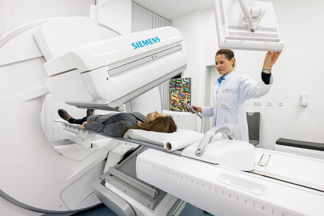
For the stress test, your heart is stressed with the help of a certain medication. You don’t have to get on a cycle ergometer. The radioactively labeled examination substance is then injected via a vein in the arm. With the blood flow, it passes through the coronary vessels into the heart muscle cells, where it is stored depending on the blood flow. Using the gamma camera, the distribution of activity in your heart muscle is imaged with what is called a scintigram.
After evaluation of the stress examination, we decide whether the so-called rest examination is necessary. For this purpose, you will be injected again with the radioactively labeled examination substance under resting conditions, and a second image of the blood flow in the heart muscle will be made by the gamma camera. By comparing the stress and rest examinations, it is possible to make accurate statements about whether you have relevant circulatory disturbances (ischemia) under stress or scarred areas after a heart attack.
The sequence of stress and rest examination can be performed in the two-day protocol, i.e. on two 2 different examination days, or on one examination day, the 1-day protocol. Advantage of the 2-day protocol is lower radiation exposure.
When is myocardial scintigraphy (especially) useful?
Warning signs of coronary artery disease, i.e., whether narrowing of the coronary arteries supplying the heart muscle is present, may include symptoms such as a squeezing sensation or burning sensation in the chest area. This may also be associated with pain radiating to the left arm or, in women, pain in the upper back between the shoulder blades. In addition, shortness of breath occurs not infrequently.
At rest, there is no discomfort in this process. However, in the case of physical stress, the above-mentioned signs occur. This is exactly where cardiac scintigraphy comes in. Through this examination method, the blood flow to the heart muscle can be examined non-invasively. Coronary artery disease is safely ruled out or detected, and your cardiologist receives important information for treatment planning.
According to the current study situation, if the cardiac scintigraphy is unremarkable and no pathological findings are detected, then the risk of a heart attack in the next five years drops to less than 1 percent. At the same time, the radiation exposure during a cardiac scintigraphy is low: it is roughly comparable to the annual natural radiation exposure in Germany.
What do you need to know before the treatment?
Fasting for cardiac scintigraphy
You must appear for the examination sober. In this way, the radioactively labeled substance can be absorbed by the heart tissue in the best possible way and accumulates only minimally in other tissue (for example, in the gastrointestinal tract).
Discontinue certain medications, no caffeine
For medicinal stress of the heart with a vasodilator, no food or drink containing caffeine (coffee, cola, black or green tea, chocolate) should be taken at least 12 h before the stress test.
Drugs containing caffeine (e.g. painkillers such as Aspirin® forte or flu drugs such as Grippostad®) and drugs containing theophylline must be paused for at least 24 h, and drugs containing dipyridamole (e.g. Aggrenox®) must be discontinued for at least 24 h.
A beta blocker should be paused 24h before (exceptions possible here in consultation with your cardiologist).
What is the procedure for a myocardial scintigraphy at Radiologie München?
Clarification interview
Before the examination, you will have a detailed informative discussion with one of our nuclear medicine specialists. During this consultation, the procedure for the examination will be explained to you, taking into account your individual medical history and complaints. In addition, your questions will be fully answered.
Load test & rest recording
At the beginning of the examination, you will be given access (arm vein). We do not perform the stress of the heart with a bicycle ergometer, but we use a drug ( = a vasodilator). This drug can trigger various symptoms, but due to the very short duration of action, the symptoms stop after about 1-2 minutes. You may find it harder to breathe. A feeling of pressure in the chest or abdomen, legs or neck, dizziness, headache or a sensation of warmth are also commonly described by patients.
After injection of the stress medication, we inject the radioactively labeled examination substance. We speak here of the load absorption. This is followed by a break of about 45 minutes. During this time you should eat a snack, drinking coffee is now allowed and you should drink at least half a liter of still water.
For the heart recordings after stress, lie on the camera for approx. 10 min. After evaluation of the images, a decision is made as to whether a second examination under resting conditions is required, the so-called resting examination. This is done either in the 1-day protocol afterwards or in the 2-day protocol usually one week later. For the rest examination, the examination substance is injected again. If you are scheduled for a 2-day protocol, please return to the appointment sober.
In the waiting time you may then eat something and please drink still water again to improve the quality of the images. Image acquisition is again performed over 10 minutes on the gamma camera. The images are evaluated, the results are discussed with you and immediately forwarded to your cardiologist.
What are the side effects of myocardial scintigraphy?
The drug exposure during myocardial scintigraphy may cause side effects such as shortness of breath, chest pain, flushing of the skin, drop in blood pressure, and cardiac arrhythmias. Strictly speaking, however, these are not side effects, but part of the effect. We will inform you in detail about the possible risks in a personal consultation before the examination.
Radiation exposure during myocardial scintigraphy is low. Depending on the examination protocol, it corresponds approximately to one to three times the annual natural radiation exposure in Germany. Decades of studies also show that radiation exposure from a scintigraphy has no health consequences.


