Scintigraphy of the lungs
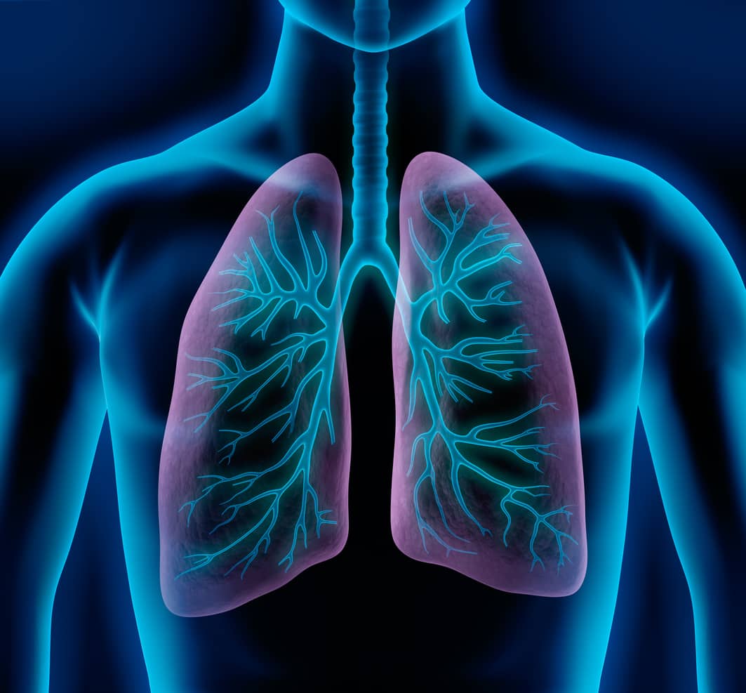
Lung scintigraphy is a nuclear medicine examination that records blood flow (perfusion) and ventilation (aeration) in the lungs. Ventilation maps the exchange of gases in the airways ( pulmonary alveoli). Perfusion shows the activity of blood flow to the small pulmonary circulation.
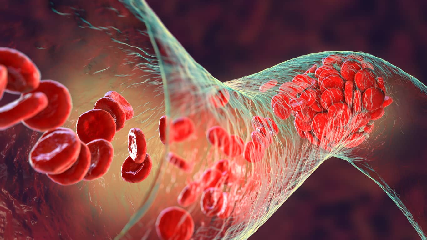
The most common diagnostic question is about pulmonary artery embolism. The loss of blood flow caused by a blood clot (embolus) can be easily visualized using perfusion scintigraphy. Thus, the embolus is localized and evaluated according to its size and number.
approx. 30 min
Duration of examination
2 to 5 times
Lower radiation exposure than with a thoracic CT scan
Possible with
Renal failure
Contrast agent allergy
Hyperthyroidism
Where can you have a lung scintigraphy performed in Munich?
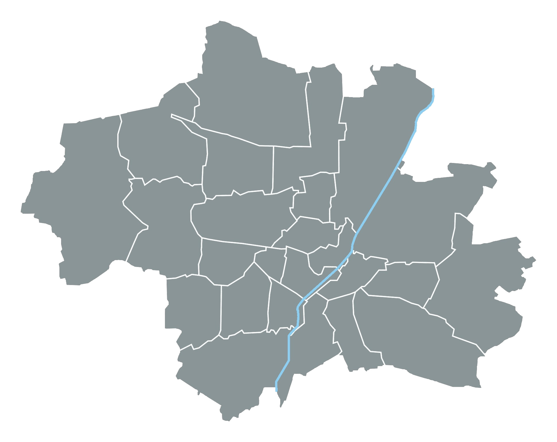
We offer perfusion scintigraphy at our location in the Rotkreuzklinikum, Nymphenburger Str. 163 in Munich (U-Bahn stop Rotkreuzplatz). Please bring any previous findings and images with you to the examination. For patients with compulsory insurance, we require a valid referral slip.
When is a lung scintigraphy useful?
Lung dysfunction is potentially life-threatening. Lung scintigraphy provides a very good diagnostic basis for various clinical pictures:
- Suspicion of acute pulmonary artery embolism
- Exclusion or detection of chronic thromboembolic pulmonary hypertension (CTEPH).
- For side-separated quantification of ventilation and perfusion preoperatively before
- Lung resection for bronchial carcinoma
- Surgical or endoscopic lung volume reduction for COPD.
- Lung Transplant
- Lung resection for non-malignant disease (e.g., localized bronchiectasis).
- Determination of a right-left shunt
How does a lung scintigraphy work?
During the ventilation examination, the radioactively labeled examination substance is inhaled (inhaled) and an image of the lung ventilation is made by the gamma camera.
In perfusion examination, the radioactively labeled examination substance (albumin = protein particles) is administered via an arm vein and its distribution in the lungs is a reflection of the blood flow. The principle here is the capillary blockade by the radioactive-labeled protein particles, which close every 10,000th capillary (0.01%) only temporarily for a short time without influencing the blood circulation. Allergy to the protein albumin used is rare.
Which radiopharmaceutical is used?
Evaporated 99m-technetium labeled carbon particles are used for the ventilation study. The particle size is very small (<0.01 µm). These are inhaled. In perfusion testing, 99mTc-labeled albumin particles are used and injected via an arm vein. The particle size is very small (diameter 15 – 40 µm).
What are the advantages of lung scintigraphy?
Pulmonary scintigraphy is also possible in cases of elevated creatinine (renal insufficiency), contrast agent intolerance, and hyperthyroidism. In these cases, CT angiography of the lungs would not be feasible.
Scintigraphy also allows the diagnosis of pulmonary artery embolism even if there is a negative result on CT angiography of the lung.
The significantly lower radiation exposure of pulmonary scintigraphy relative to CT angiography offers several additional applications. Women, including those during pregnancy and after delivery, can be examined. The same applies to younger patients, regardless of their gender.
What do you need to know before the treatment?
You do not have to be sober. Please bring any previous findings and previous images / CT scan of the lungs with you to the examination. No magnesium sulfate infusion may have taken place beforehand and please inform us if you are taking chemotherapeutic agents, as certain medications may be affected.
What is the procedure for a lung scintigraphy at Radiologie München?
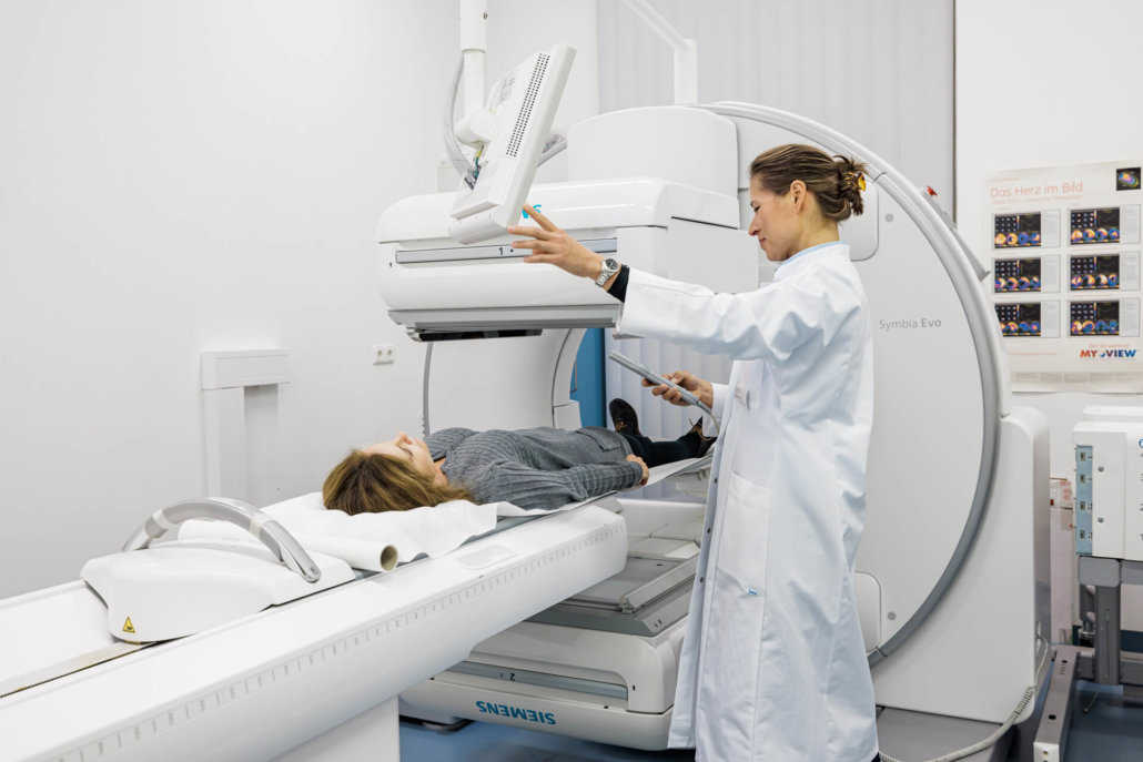
For the ventilation examination, you breathe the examination substance deep into the bronchi of the lungs via a mask. Drinking water and rinsing the mouth will eliminate any remaining activity in the mouth and esophagus. They then lie on the gamma camera for 20 min, which slowly rotates around them and images the ventilation of the lungs through the activity distribution.
During the perfusion examination, you lie on the gamma camera. We ask them to inhale and exhale deeply several times that the lungs can develop well. The test substance consisting of 99m-technetium-labeled albumin particles is injected via an arm vein. The recordings also take about 20 min and map the blood flow to the lungs through the activity distribution.
Our nuclear medicine physician will discuss the findings with you following the examination.
What are the risks and side effects of lung scintigraphy?
A suspension solution of human albumin particles is used as the test substance. An allergy to albumin (= protein) is rare. It must not be used if you are hypersensitive (allergic) to it. There are isolated reports of hypersensitivity reactions or local allergic reactions at the injection site.
