X-ray digital
X-ray diagnostics (digital radiography) is still very important at present due to its fast and cost-effective method. As a diagnostic tool or as part of the overall diagnosis, digital radiography is used to clarify diseases of the lungs, bones or airways.
Radiography often provides the first clues for the detection of a disease, which are clarified in more detail in further examinations such as CT or MRI.
approx. 1-2 min
Duration of the examination
approx. 30 min
Stay in the practice
Your digital radiography at Radiologie München
We offer X-ray screening at all of our radiology locations. Thanks to state-of-the-art technology, the doses of X-rays used are kept extremely low. Since the images are provided in digital form, your treating medical team can view the results quickly and easily on the digital patient portal.

How does digital radiography work?
X-ray imaging still works according to the well-known principle, in which a specific area of the human body is illuminated directionally with X-rays. The X-rays strike a very highly reactive medium behind the body and image the different densities of tissue and bone in varying shades.
In digital radioraphy, the earlier X-ray films are replaced by sensor plates that transmit the image directly to the examination computer. This makes it possible to produce more precise images of very well delimited areas. This also significantly reduces the radiation exposure during the examination.
Where does digital radiography offer the best diagnostic opportunities?
Radiographs are particularly useful for diagnosing fractures and wear and tear diseases of the spine. This is due to the very good representability of the bone apparatus in the radiographic image. The X-ray images can then be used for immediate treatment or for regional containment of further investigative methods.
In the area of the lung, there are various areas of application, in particular for the detection of
- Pneumonia,
- Lung tumors,
- pleurisy or
- of pulmonary congestion in heart failure.
In addition, digital radiography is used for the diagnosis of breast cancer (mammography).
What should you consider before the examination?
Before the examination, all foreign bodies that could interfere with radiography should be removed. In particular, piercings and body jewelry should be removed from the areas to be examined. We recommend that you remove all metal objects such as watches, jewellery, hair clips or the underwire bra beforehand.
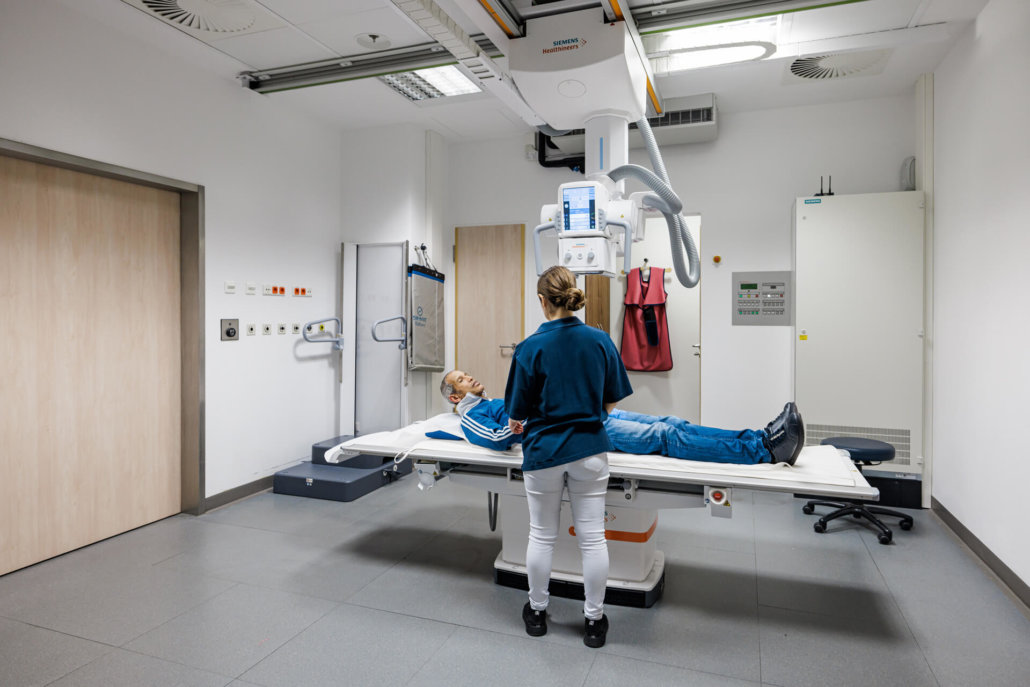
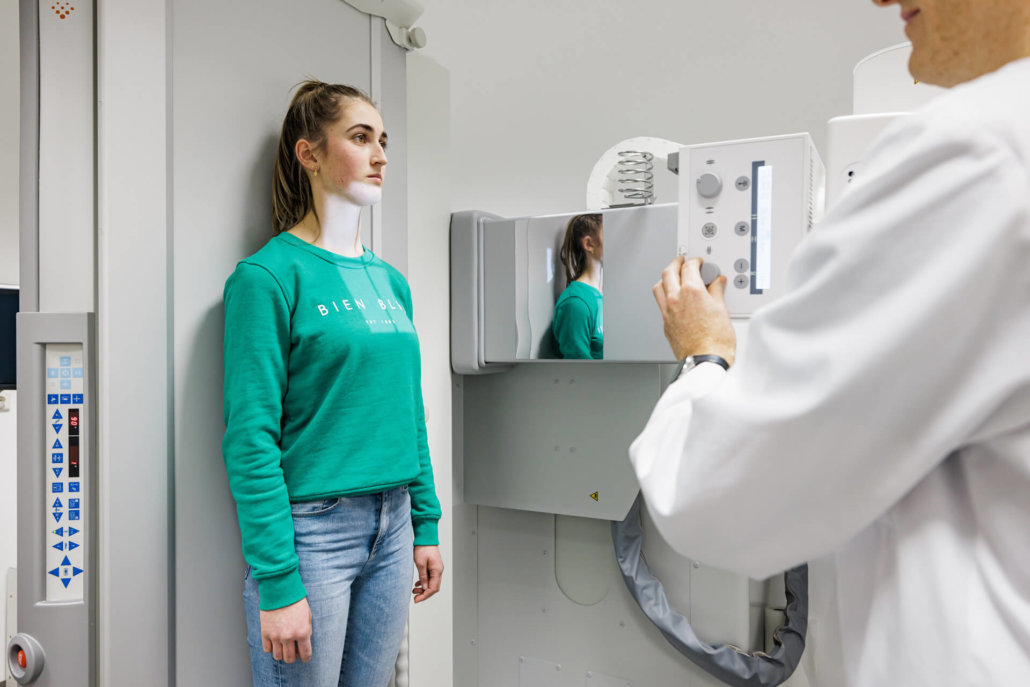
You will be placed in the necessary position by our trained, friendly staff before the examination. Particularly radiation-sensitive regions can be protected by lead aprons.
During the examination, you must try to lie perfectly still to avoid blurring during image acquisition. Thanks to digital radiography, the images are available directly after the examination during the consultation with one of our specialists and can thus be evaluated very quickly.


