CT CONTROLLED
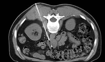
BIOPSY / DRAINAGE
CT CONTROLLED
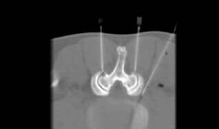
PAIN THERAPY
Interventional radiology
As an equally innovative and sophisticated subfield of diagnostic radiology, interventional radiology uses imaging examination procedures to perform targeted and highly precise therapeutic and diagnostic interventions.
We perform the vast majority of image-guided procedures CT-guided. Typical procedures include tumor ablation using microwave or radiofrequency ablation, and drainage to remove local inflammation or pathological fluid collections. Local pain therapy, for example in the case of an acute herniated disc (PRT), is also one of the areas of application.
No matter which procedure is used in interventional radiology – the advantages for our patients are manifold: they are minimally invasive interventions, i.e. extremely gentle for you, and can therefore usually be performed on an outpatient basis. As a rule, we can perform the procedure without anesthesia and only under local anesthesia. For you, this means that you can already go home after only a short stay in the hospital.
In principle, many procedures are performed equally by our entire team of doctors, but for more complex issues you are always welcome to contact Prof. Herzog or Prof. Mack.
CT-guided biopsy & drainage
CT-guided biopsy is used when an unclear mass (suspected tumor, inflammatory changes, etc.) requires minimally invasive tissue collection for subsequent histological or microbiological examination.
CT-guided pain therapy
In pain management, more accurate and effective treatment outcomes can be achieved using CT-assisted measures. The following types of pain therapy may be considered:
- Periradicular therapy (PRT)
- Facet infiltration
- Epidural flooding
Preliminary talk & contrast agent administration
At Radiologie München, every examination is preceded by a detailed preliminary discussion. Our specialists will explain the rest of the computed tomography procedure and the effects of the contrast agent used. In this conversation, you can express your fears about the examination. In the further procedure, a contrast agent containing iodine would be injected via the arm vein. The examination takes place in the supine position following the injection.
After the examination
You will remain for a period of time for outpatient monitoring. In the case of biopsies, the removed tissue is sent to the appropriate laboratory for examination, and you will then receive the results from your attending physician(s). In the case of drains, a final discussion takes place on site about the further procedure and necessary behavior. In the case of multiple therapies, further treatments follow according to the procedures agreed with our specialists.
What are the risks of CT-guided intervention?
In addition to the respective risks of each intervention therapy, there is a small amount of radiation exposure and the injection of an iodine-containing contrast agent, just as with a conventional CT scan.
Radiation exposure risk
The CT scan involves radiation exposure to the body, which is higher than normal X-ray examinations. The amount of exposure depends on the dose administered, the duration of the examination, and the tissue being examined. In any case, dual-energy computed tomography should only be performed if it is needed by the doctor to detect and type certain diseases.
Risk contrast agent
The contrast agent may cause headache, dizziness, nausea, diarrhea or abdominal pain in some cases. Here it is recommended to drink a lot of water after the treatment. If there is no improvement in the near future, you must contact us immediately. Allergic reactions can also occur unexpectedly, but these can be treated with medication.


