What does neuroradiology mean?
The field of neuroradiology includes diagnostics and therapy for pathological changes in the brain and spinal cord.
The methods of neuroradiology support not only neurology and neurosurgery, but also ENT, ophthalmology and maxillofacial surgery.
Arrange online appointment
Quickly and easily get an appointment at a Radiologie München practice in Radiologie München.
Radiologie München – our locations
For your neuroradiological examination, Radiologie München has a variety of locations available in the Munich metropolitan area. In the north of Munich, we also have a practice in Unterschleißheim.
Pick a location that works for you and make an appointment.

Range of services
Neuroradiology uses digital radiography (X-ray examination), CT and MRI, as well as sonography and myelography (examination of the spinal canal with contrast medium).
Important information can also be gathered by angiography using MRI or CT. Feel free to find out more about it via the following link.
CT of the brain and spine
Computed tomography, or CT, creates detailed images of the brain and spine. In neuroradiology, head CT is often used for acute issues, such as clarifying a possible brain hemorrhage, which is always a medical emergency. Furthermore, if a stroke is suspected, the blood flow to certain areas of the brain can be examined in detail – a decisive basis for further therapy.
A CT, unlike an MRI, uses low-dose X-rays. During the examination, the device automatically moves you – usually lying on your back – through the extra large opening of the detector. Software is used to calculate the images and play them out digitally in very thin slices.
Under certain conditions, a contrast agent containing iodine may be used. Its injection usually leads to a feeling of warmth. However, this is not bad and disappears after a few minutes. Apart from this feeling of warmth, the contrast medium is usually very well tolerated. In the case of impaired kidney function or problems with the thyroid gland, the contrast medium tolerance should be checked beforehand by our specialists.
MRI scans of the brain and spinal cord
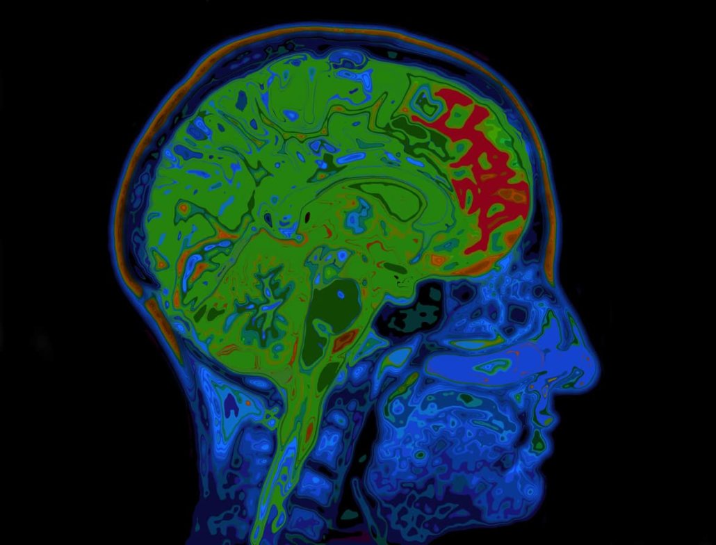
A head MRI is used to obtain very detailed slice images of the brain and spinal cord. Due to the excellent contrast of the images, strokes can be reliably diagnosed a very short time after the first symptoms.
In addition, an MRI produces images of the vessels in the brain and allows temporal processes of blood flow to be visualized. The advantage of head MRI is that neither X-rays (but magnetic fields) affect the head, nor contrast media containing iodine are used.
If a contrast medium is used in rare cases, it is a particularly low-risk, macrocyclic contrast medium that is very well tolerated. If your kidney function is severely restricted, please let us know in good time so that we can clarify the tolerance of the contrast medium in advance.


